Review
Similar Products
|
ATCC
human osteosarcoma mg63 cell line Human Osteosarcoma Mg63 Cell Line, supplied by ATCC, used in various techniques. Bioz Stars score: 98/100, based on 1 PubMed citations. ZERO BIAS - scores, article reviews, protocol conditions and more https://www.bioz.com/result/human osteosarcoma mg63 cell line/product/ATCC Average 98 stars, based on 1 article reviews
human osteosarcoma mg63 cell line - by Bioz Stars,
2026-06
98/100 stars
|
Buy from Supplier |
|
Procell Inc
human osteosarcoma cell line mg63  Human Osteosarcoma Cell Line Mg63, supplied by Procell Inc, used in various techniques. Bioz Stars score: 86/100, based on 1 PubMed citations. ZERO BIAS - scores, article reviews, protocol conditions and more https://www.bioz.com/result/human osteosarcoma cell line mg63/product/Procell Inc Average 86 stars, based on 1 article reviews
human osteosarcoma cell line mg63 - by Bioz Stars,
2026-06
86/100 stars
|
Buy from Supplier |
|
ATCC
human osteosarcoma cell line mg63  Human Osteosarcoma Cell Line Mg63, supplied by ATCC, used in various techniques. Bioz Stars score: 98/100, based on 1 PubMed citations. ZERO BIAS - scores, article reviews, protocol conditions and more https://www.bioz.com/result/human osteosarcoma cell line mg63/product/ATCC Average 98 stars, based on 1 article reviews
human osteosarcoma cell line mg63 - by Bioz Stars,
2026-06
98/100 stars
|
Buy from Supplier |
|
ATCC
human osteosarcoma cell lines mg63  Human Osteosarcoma Cell Lines Mg63, supplied by ATCC, used in various techniques. Bioz Stars score: 98/100, based on 1 PubMed citations. ZERO BIAS - scores, article reviews, protocol conditions and more https://www.bioz.com/result/human osteosarcoma cell lines mg63/product/ATCC Average 98 stars, based on 1 article reviews
human osteosarcoma cell lines mg63 - by Bioz Stars,
2026-06
98/100 stars
|
Buy from Supplier |
|
JCRB Cell Bank
human osteosarcoma cell line mg63 Human Osteosarcoma Cell Line Mg63, supplied by JCRB Cell Bank, used in various techniques. Bioz Stars score: 90/100, based on 1 PubMed citations. ZERO BIAS - scores, article reviews, protocol conditions and more https://www.bioz.com/result/human osteosarcoma cell line mg63/product/JCRB Cell Bank Average 90 stars, based on 1 article reviews
human osteosarcoma cell line mg63 - by Bioz Stars,
2026-06
90/100 stars
|
Buy from Supplier |
|
ATCC
mg63 human osteosarcoma cell line 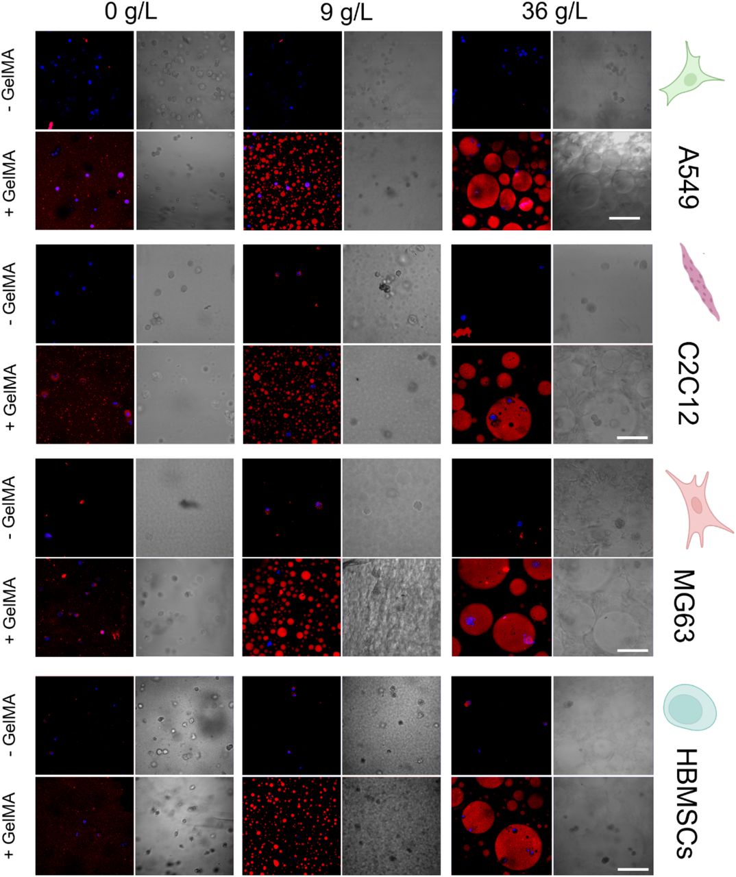 Mg63 Human Osteosarcoma Cell Line, supplied by ATCC, used in various techniques. Bioz Stars score: 98/100, based on 1 PubMed citations. ZERO BIAS - scores, article reviews, protocol conditions and more https://www.bioz.com/result/mg63 human osteosarcoma cell line/product/ATCC Average 98 stars, based on 1 article reviews
mg63 human osteosarcoma cell line - by Bioz Stars,
2026-06
98/100 stars
|
Buy from Supplier |
|
ATCC
human cell line mg63 osteosarcoma Human Cell Line Mg63 Osteosarcoma, supplied by ATCC, used in various techniques. Bioz Stars score: 98/100, based on 1 PubMed citations. ZERO BIAS - scores, article reviews, protocol conditions and more https://www.bioz.com/result/human cell line mg63 osteosarcoma/product/ATCC Average 98 stars, based on 1 article reviews
human cell line mg63 osteosarcoma - by Bioz Stars,
2026-06
98/100 stars
|
Buy from Supplier |
Image Search Results
Journal: RSC Advances
Article Title: Replicating the post-chemotherapy tumor microenvironment via biomimetic scaffolds to regulate MSC differentiation
doi: 10.1039/d6ra01031h
Figure Lengend Snippet: Biological characterizations of DOX-MSNCaP@PLGA-collagen and PLGA-collagen constructs. The accumulate release of DOX from DOX-MSNCaP@PLGA-collagen in PBS buffer with different pH (A). Cytotoxic assessment of MG63 cells after being cultured in scaffolds via live and dead staining. Cells were seed in the meshes and incubated in DMEM at pH 7.4 or pH 6.5 (B). Fluorescence microscopic observation on the intracellular distribution of DOX in MG63 cells after incubation with DOX-MSNCaP@PLGA-collagen scaffolds for 12 hours. Cell nucleus was represented blue fluorescence; DOX was represented red fluorescence; actin cytoskeleton was represented green fluorescence (C). DNA laddering analysis on the severity of DNA fragmentation in MG63 cells upon treatment of DOX-MSNCaP@PLGA-collagen for 12 hours (I: PLGA-collagen at pH 7.4, II: DOX-MSNCaP@PLGA-collagen at pH 7.4, III: DOX-MSNCaP@PLGA-collagen at pH 6.5) (D).
Article Snippet: Different types of cells were utilized in this work, including human bone marrow-derived mesenchymal stem cells (MSCs, passage 4) obtained from LONZA (MD, USA), and the
Techniques: Construct, Cell Culture, Staining, Incubation, Fluorescence, DNA Laddering

Journal: RSC Advances
Article Title: Replicating the post-chemotherapy tumor microenvironment via biomimetic scaffolds to regulate MSC differentiation
doi: 10.1039/d6ra01031h
Figure Lengend Snippet: BrdU staining of MG63 upon treatment of DOX-MSNCaP@PLGA-collagen at pH 7.4 or pH 6.5. Scale bar: 50 µm.
Article Snippet: Different types of cells were utilized in this work, including human bone marrow-derived mesenchymal stem cells (MSCs, passage 4) obtained from LONZA (MD, USA), and the
Techniques: BrdU Staining
Journal: Polymers
Article Title: Localized Combination Therapy Using Collagen–Hydroxyapatite Bone Grafts for Simultaneous Bone Cancer Inhibition and Tissue Regeneration
doi: 10.3390/polym17162239
Figure Lengend Snippet: Cytotoxicity effect on MG63 ( a ) and BMSCs ( b ) after 24 h of exposure to the tested samples’ extracts, evaluated via the release of LDH enzyme. Complete growth media was used as a negative control, and lysed cells were used as a positive control. Results are expressed as mean ± standard deviation (n ≥ 3, independent samples).
Article Snippet: For cell culture,
Techniques: Negative Control, Positive Control, Standard Deviation

Journal: Polymers
Article Title: Localized Combination Therapy Using Collagen–Hydroxyapatite Bone Grafts for Simultaneous Bone Cancer Inhibition and Tissue Regeneration
doi: 10.3390/polym17162239
Figure Lengend Snippet: Cellular viability of MG63 ( a ) and BMSCs ( b ) cultured with 100% extracts from each sample, for 1 day. Complete growth media was used as a control. The dashed line indicates the 70% cut-off for a toxic effect. Results are expressed as mean ± standard deviation (n ≥ 3, independent samples, * p < 0.05).
Article Snippet: For cell culture,
Techniques: Cell Culture, Control, Standard Deviation

Journal: Polymers
Article Title: Localized Combination Therapy Using Collagen–Hydroxyapatite Bone Grafts for Simultaneous Bone Cancer Inhibition and Tissue Regeneration
doi: 10.3390/polym17162239
Figure Lengend Snippet: Cellular viability of MG63 ( a ) and BMSCs ( b ) cultured with 100% extracts from each sample for 3 days. Results are expressed as mean ± standard deviation (n ≥ 3; independent samples; * p < 0.05).
Article Snippet: For cell culture,
Techniques: Cell Culture, Standard Deviation
Journal: Molecular Nutrition & Food Research
Article Title: Saucerneol Inhibits the Growth, Migration, and Invasion of Osteosarcoma Cells In Vitro and Prevents Metastasis‐Associated Osteolysis Ex Vivo
doi: 10.1002/mnfr.70187
Figure Lengend Snippet: Effects of Sauc on cell death, apoptotic cell morphology, and migration in human osteosarcoma cells. (A, B) MG63 cells (A) and SJSA‐1 cells (B) were seeded in 96‐well plates and cultured with Sauc at the indicated concentrations for 24 h. The numerical values of cytotoxicity are presented as a bar graph normalized to those of the control. (C, D) MG63 cells (C) and SJSA‐1 cells (D) were seeded in six‐well plates and cultured with Sauc at the indicated concentrations for 24 h. The morphological changes were observed using the Olympus CKX53 inverted microscope. Scale bar: 200 µm. (E, F) After 24 h of Sauc treatment of MG63 cells (E) and SJSA‐1 cells (F), cell migration was assessed using the wound healing assay. The migration rate is presented as a bar graph. Scale bar: 200 µm. All values are expressed as mean ± SD of the results from three independent experiments. * Indicates a statistically significant difference, with p < 0.05 compared to the control.
Article Snippet:
Techniques: Migration, Cell Culture, Control, Inverted Microscopy, Wound Healing Assay
Journal: bioRxiv
Article Title: Aqueous two-phase bioinks for discrete packing and compartmentalisation of 3D bioprinted cells
doi: 10.1101/2025.06.27.661968
Figure Lengend Snippet: Partitioning of different cell types within the various ATPS scaffold formulations. Brightfield and confocal (merged) images representative of cells labelled with DAPI to show the nucleus (blue) of the different cell types (A549, C2C12, MG63, HBMSCs), and GelMA marked with rhodamine B to show the inner phase (red) of the scaffolds at 0 - 9 - 36 g/L. In all cell types, images were taken both under conditions where GelMA was not chemically cross-linked (-GelMA) and under conditions where GelMA was chemically cross-linked (+GelMA). Scale bars: (a, b, c, d) 100 μm. Mean ± S.D. n=3.
Article Snippet: The A549 human lung carcinoma epithelial-like cell line, C2C12 mouse pre-myoblast cell line and
Techniques: